Usefulness of new imaging methods for assessment of type B aortic dissection
Abstract
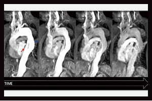
While the medical management of uncomplicated type B aortic dissection has good outcomes in the short term, the longer term mortality can be in the region of 50% at 5 years. Up to 40% of the survivors can have significant dilatation of the false lumen with the risk of aneurysm formation and death due to rupture. The results of the randomized controlled trials ADSORB and INSTEAD-XL have shown that beneficial aortic remodelling occurs after endoluminal stent graft placement, but these trials were underpowered to show any effect on survival. Static computed tomography (CT) angiography imaging methods have been used to try to identify high risk patients using parameters such as diameter, the position and size of the entry tear, and the amount of false lumen thrombus, but these so far are not able to clinically risk stratify individual patients. In this manuscript, we present our initial experience with new MR imaging methods. These have allowed us to develop a greater understanding of aortic dissection by providing information regarding the underlying hemodynamic and biomechanics of the dissection, as well as more accurate assessment of important clinical imaging endpoints, such as false lumen thrombosis.
Cover


