Article Abstract
Techniques to define segmental anatomy during segmentectomy
Abstract
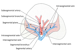
Pulmonary segmentectomy is generally acknowledged to be more technically complex than lobectomy. Three-dimensional computed tomography (3D CT) angiography is useful for understanding the pulmonary arterial and venous branching, as well as planning the surgery to secure adequate surgical margins. Comprehension of the intersegmental and intrasegmental veins makes the parenchymal dissection easier. To visualize the segmental border, creation of an inflation-deflation line by using a method of inflating the affected segment has become the standard in small-sized lung cancer surgery. Various modifications to create the segmental demarcation line have been devised to accurately perform the segmentectomy procedure.
Cover


